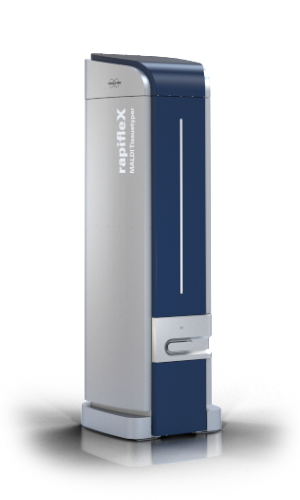
rapifleX MALDI Tissuetyper The rapifleX® MALDI Tissuetyper® re-defines the key performance measures for MALDI Imaging
Tissue typing – A clinical discovery workflow
The speed of the rapifleX® Tissuetyper® allows for fast acquisition of images and SCiLS™ Lab solution seamlessly allows for classification of the data sets, enabling tissue typing at minimal time. Tissue micro arrays can be analyzed very rapidly. The soft smartbeam 3D laser effectively ionizes the analytes but does not harm the tissue, in fact histological staining is possible after the acquisition of the MALDI data set.
The combination of the mass spectrometric, molecular information with the histology of the analyzed tissue yields information-rich data sets, resulting in deeper understanding of the spatial distribution of molecules linked to the observed biology. The image displays a typical workflow, and also shows some statistical features of the SCiLS™ Lab solution
 Inquery Form